

The purpose of mole removal is to completely eliminate the mole from the body. This can be achieved either by surgical methods, which involve cutting out the nevus, or by non-surgical methods, such as vaporization or ‘mole burning’, which do not require cutting.
Non-surgical mole removal methods include laser, radiofrequency, plasma, electrosurgery, and cryotherapy (freezing the moles), the latter being the most commonly applied by dermatology specialists. Additionally, moles can naturally decrease in size and disappear as a result of aging.
Which is the Best Treatment Method?
The answer to this question may vary from one doctor to another. A doctor who performs surgical mole removal may say, “Surgery is the most effective method.” Another might claim, “Laser treatment for moles is the best.” For me, the best method is “Radiofrequency.” Here, what matters is the experience and skill of the practitioner.
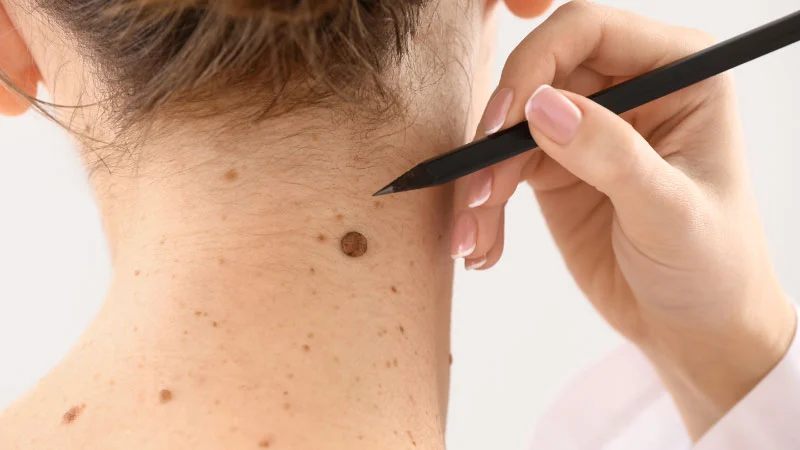
How is Mole Removal with Radiofrequency Performed?
Radiofrequency is a method based on radio (sound) waves, used for vaporizing lesions such as moles, warts, and calluses. Although similar to electrosurgery, radiofrequency specifically targets the treated area without damaging surrounding tissues.
What are the Advantages of Radiofrequency Treatment?
Compared to other methods, such as laser, plasma, and electrosurgery, radiofrequency can remove all types of moles. Superficial, raised, colored, or light-colored moles can all be treated in minutes without needing another treatment method.
For more images and videos, I recommend following the ‘Mole Removal Instagram’ channel where before and after pictures of mole removal procedures are shared.
How Long is the Healing Process After Mole Removal?
Initially, the treated area will form a scab resembling a wound. This scab will then give way to pink skin tissue, which will return to its normal color over time. This process can vary from 10 days to a month, depending on the individual’s skin condition.
Will There Be a Scar After Mole Removal?
Based on my own experience, there is usually no significant scarring following mole removal with radiofrequency. In the initial application, we perform a superficial procedure without going too deep to avoid scarring. If there is still some swelling or darkness after two months, a deeper (free touch-up) procedure can be performed.
During the healing process, temporary scabbing and a pinkish appearance on the skin will occur.
Should Cream Be Used After Mole Removal?
Although not absolutely necessary, creams like Kutalin can be used to accelerate the healing process.
What are Moles?
Mole types are classified according to their origins (whether they are congenital or acquired), as well as their colors and sizes. We will provide a summary of the most common types of moles found in Turkey.
Junctional Nevus: This condition arises when cells destined to form a nevus develop in the initial stage between the upper layer of the skin, the epidermis, and the middle part, the dermis. They appear flat and pigmented.
Compound Nevus: This type occurs when nevus cells spread into the dermis (deeper layers).
Dermal Nevus: Some moles are found solely in the dermis. They are usually raised above the surface of the skin. Those that are colorless are known as cellular nevi.
Blue Nevus: These are deep blue in color.
Pigmented Nevus (Congenital): These are dark-colored moles present from birth and can vary in size. They can cover areas larger than the size of a pinhead. Especially large ones should be carefully monitored for the possibility of turning cancerous.
Halo Nevus: Some moles are surrounded by a white ring. These types of nevi are usually seen in childhood or adolescence and are harmless. They may disappear over time.
Freckles: These are light brown, usually small, and not raised above the skin. They are common on body parts exposed to the sun and in people who are exposed to the sun. Their color darkens with sun exposure and fades during winter months. They are more common in people with fair skin and are often confused with moles.
Sunspots: The color of some sunspots may darken with sun exposure and may not lighten in winter. These spots can rarely turn into cancer and are known as Lentigo solaris.
Atypical Nevus: Also known as Clark Nevus, this type of mole is considered to have a risk of turning into cancer due to its irregular borders and large size. However, most biopsies performed are unnecessary, and the results are usually benign. If there is a family history of melanoma, careful monitoring and possibly a biopsy are recommended in these cases.
Are Moles Harmful?
The vast majority of moles found on the skin are benign. They usually do not grow or change. However, during puberty and pregnancy, the number of moles can increase, or they may grow slightly.
Some moles can become malignant; for example, they can turn into a skin cancer such as malignant melanoma. The probability of moles becoming malignant is, on average, one in 200,000. People with moles larger than 6 mm should be cautious.
Moles known as dysplastic nevi range in size from 5 to 30 mm and are generally larger than other moles that form later. They can appear on different parts of the body but are most commonly found on the torso. Special attention should be paid to these moles, particularly in individuals who develop them between the ages of 20 and 30 and those with a family history of widespread moles, especially if there is a history of melanoma. In the presence of familial melanoma, the rate of these moles becoming cancerous is much higher. In those without a family history, the cancer rate is much lower.